In the ongoing study of viruses, researchers are using powerful computer resources to create visualizations of virus structures. MSI Principal Investigator Wei Zhang, a research assistant professor in the Department of Diagnostic and Biological Sciences (School of Dentistry), is investigating several viruses by using cryoelectron microscopy and creating three-dimensional reconstructions of their structures using advanced software.
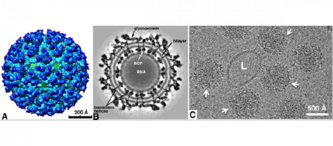
In a recent paper that appeared in the Proceedings of the National Academy of Sciences of the USA, Professor Zhang and Research Associate Dr. Sheng Cao described how they were able to create visualizations of an intermediate step in membrane fusion, using the Sindbis virus, a prototypical alphavirus. (Sheng Cao and Wei Zhang. 2013. Characterization of an early-stage fusion intermediate of Sindbis virus using cryoelectron microscopy. Proceedings of the National Academy of Sciences of the United States of America 110 (33) (AUG 13): 13362-7.) The authors created an environment where the virus could bind with liposomes, took electron micrographs of the virus-liposome complex, and generated three-dimensional reconstructions. These 3D images show the structure of an early-stage fusion intermediate of an enveloped virus and may be beneficial in determining target locations for antiviral drugs. The authors have placed the reconstructions in the EMDataBank (www.emdatabank.org), an online resource for storing and sharing electron microscopy images.
Professor Zhang also uses MSI resources to study the human T-lymphotropic virus type 1 (HTLV-1), which is responsible for a number of disorders including adult T cell leukemia/lymphoma and HTLV-1 associated myelopathy/tropical paraperesis. As they did with the Sindbis virus, the Zhang group uses cryoelectron microscopy and three-dimensional reconstruction to study the structure of HTLV-1. This work may provide insights that will help in drug design.
Image description: Cryo-EM image and reconstruction of Sindbis viruses (SINV). (A-B) 3D reconstruction and cross-section of SINV (Zhang, W., S. Mukhopadhyay, S. V. Pletnev, T. S. Baker, R. J. Kuhn, M. G. Rossmann. 2002. Placement of the structural proteins in Sindbis virus. J. Virol. 76:11645-11658). (C) Image of the SINV viral particles (arrows) attached to a liposome (L) at acidic pH (Sheng Cao and Wei Zhang. 2013. Characterization of an Early-Stage Fusion Intermediate of Sindbis Virus using Cryo-electron Microscopy. PNAS 110: 13362-13367).
posted July 9, 2014